Figure 3.
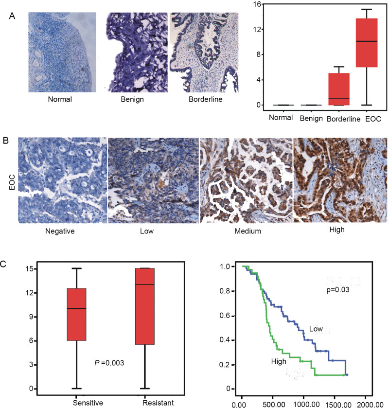
Expression of HSP27 is associated with cisplatin response in clinical samples (A, B) Immunohistochemical analysis of sections from paraffin-embedded blocks of tissues. Sections were stained with antibodies directed against HSP27 in normal and benign tissue or borderline tumor and (EOC). Anti-HSP27 antibody for immunohistochemical (IHC) analysis with scrambled probe and phosphate-buffered saline as a negative control, respectively. Representative photographs are shown. The differences of HSP27 expression between chemotherapy-sensitive and -resistant patients in ovarian cancer specimens is shown (p = 0.03, Mann–Whitney U test). (C) A Kaplan–Meier analysis of progression-free survival (PFS) for ovarian cancer patients with the corresponding expression profiles of HSP27 is shown (p = 0.03 and p = 0.01, respectively, log-rank test). All statistical tests were two sided.

