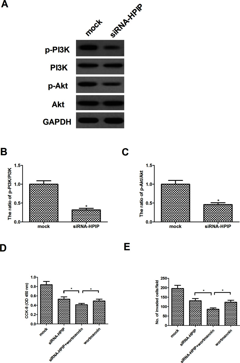
Figure 4.
HPIP silencing inhibits the activation of PI3K/Akt signaling pathway in HNSCC cells. HSC-3 cells were transfected with siRNA-HPIP or mock for 24 h. (A) The levels of phosphorylated PI3K, total PI3K, phosphorylated Akt, and total Akt were detected by Western blot analysis. Quantification of (B) p-PI3K/PI3K and (C) p-Akt/Akt. *p < 0.05 versus mock group. (D) HSC-3 cells were transfected with siRNA-HPIP or mock in the presence or absence of the wortmannin (100 nM) for 24 h. Cell proliferation was detected by the CCK-8 assay. (E) Cell invasion was evaluated by the Transwell invasion chamber assay. The results are expressed as mean ± SD and n = 3 per group. *p < 0.05.

