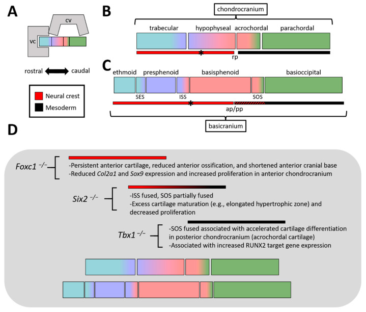
Figure 3.
(A) (Top) Block schematic of viscerocranium, calvaria, and chondro/basicranium. (Bottom) Key for panels B, C, and D, highlighting orientation of rostral-caudal axis and neural crest—mesoderm color scheme. (B) Basic diagram of chondrocranial elements (along a midsagittal plane), highlighting the crisp neural crest—mesoderm interface at Rathke’s pouch (rp). The exception to this boundary is the hypochiasmatic cartilages (denoted by asterisk), which are mesoderm derived. (C) Same as B, but highlighting elements found within the basicranium (similar to Figure 2, just schematized). Note, the shades of blue are meant to provide a relative reference to their chondrocranial precursors, although boundaries are not meant to be exact. Also highlighted are the synchondroses (e.g., SES, ISS, SOS). The neural crest—mesoderm interface is found near the anterior-posterior pituitary (ap/pp), although this interface is substantially intermixed (indicated by red/black hash) within the basicranium. (D) Summary of the phenotypes in the 3 loss-of-function models covered in this review and where defects are located relative to the cranial base. Abbreviations: ap, anterior pituitary; cv, calvaria; ISS, inter-sphenoid synchondrosis; pp, posterior pituitary; rp, Rathke’s pouch; SOS, spheno-occipital synchondrosis; vc, viscerocranium.

