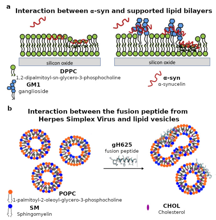
Figure 3.
(a) Schematic representation of the interaction between -synuclein (-syn) and supported lipid bilayers composed of either pure 1,2-dipalmitoyl-sn-glycero-3-phosphocholine (DPPC) or a mixture of DPPC and ganglioside GM1. Neutron reflectometry measurements revealed that GM1 affects the location of -syn with respect to the biomimetic lipid membrane. Adapted and reprinted with permission from [97], Copyright (2019) Elsevier. (b) Schematic representation of the interaction between the gH625 fusion peptide from the envelope of the herpes simplex virus (HSV) and lipid vesicles with lipid composition mimicking the rafts in the mammalian plasma membrane. Electron paramagnetic spectroscopy measurements revealed that the peptide mainly interacts with the lipid headgroups. Adapted and reprinted with permission from [113], Copyright (2015) Royal Society of Chemistry (RSC).

