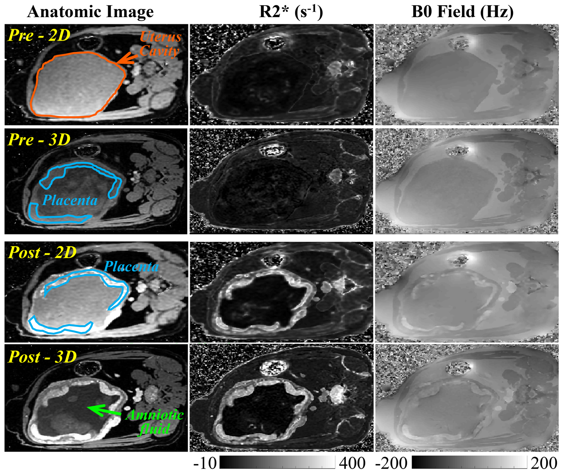
Figure 3.

Representative anatomic images, R2* maps, and B0 field maps from 2D and 3D CSE-MRI acquisitions in a pregnant rhesus macaque (Rhesus #4) at scans before (Pre) and immediately after (Post) ferumoxytol administration. Two placental discs and the uterus cavity are delineated by blue lines and orange lines, respectively. The amniotic fluid is indicated with a green arrow on the anatomic image.
