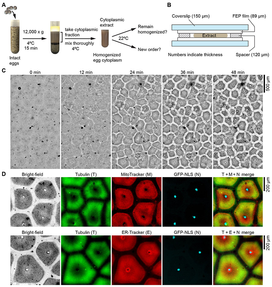
Fig. 1. Homogenized Xenopus laevis egg extracts self-organize into cell-like compartments.
(A) Schematic diagram of experimental procedures. (B) The design of the chamber used to image extracts. (C) Bright-field microscopy showing that homogenized Xenopus laevis egg cytoplasmic extracts spontaneously organized into cell-like compartments in a multi-millimeter scale field. Pattern formation dynamics in bright-field, tubulin and ER channels are presented in movie S1 and S2. (D) Spatial organization of microtubules, endoplasmic reticulum, nuclei and mitochondria in the cell-like compartments, visualized by added HiLyte 647 labeled porcine tubulin, ER-Tracker Red, GFP-NLS, and MitoTracker Red CMXRos. Pattern formation dynamics are presented in movie S3. The still images shown in (D) are from the last frame of movie S3, at 60.6 min after imaging began.

