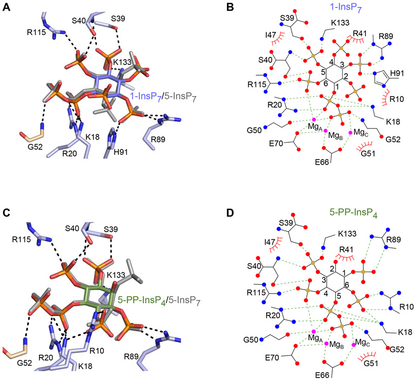
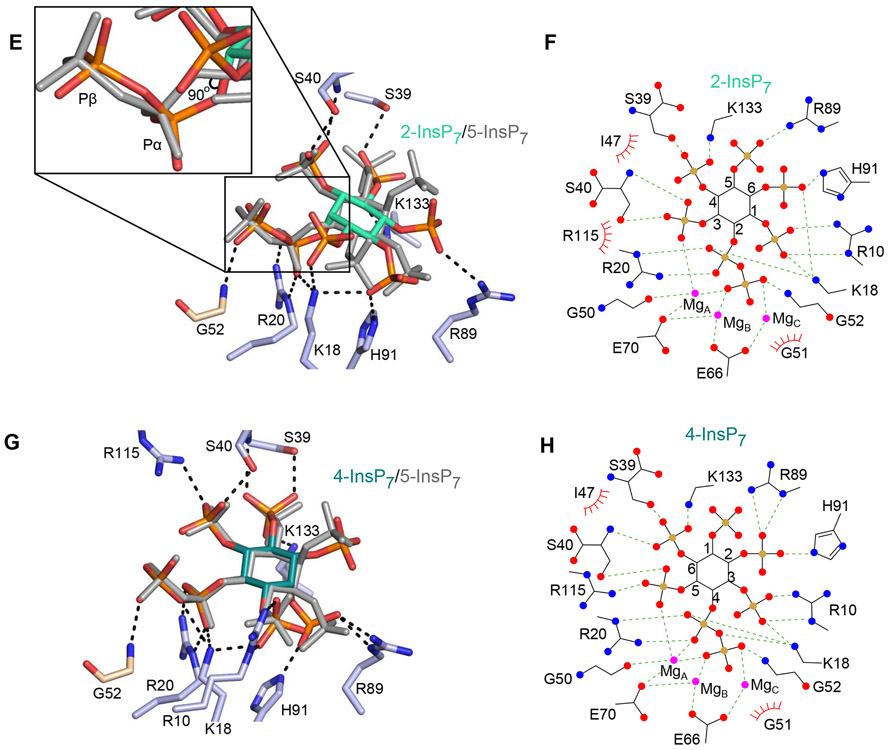
Figure 5.
DIPP1 binding of multiple PP-InsP ligands. Descriptions of amino acid residues (stick format) that have polar interactions with the indicated PP-InsP (< 3.2 Å; broken lines). All data were obtained by either co-crystallization of protein and ligand, or by ligand soaking into preformed protein crystals. Residues belonging to the Nudix motif are wheat-colored. Nitrogen is colored blue, oxygen is red and phosphorous is orange. Inositol ring carbons are color-coded as described below; the positions of each PP-InsP ligand are shown relative to 5-InsP7, which is depicted in light gray. Corresponding color-colored ligplots describe interactions between DIPP1, Mg (colored magenta), and the indicated PP-InsP; inositol carbons are numbered, polar contacts are denoted as broken lines, and Van der Waals contacts are shown as “eyelash” graphics. A, 1-InsP7 (slate blue carbons) and B, corresponding ligplot. C, 5-PP-InsP4 (avocado green carbons) and D, corresponding ligplot. E, 2-InsP7 (turquoise carbons), with magnified inset to 90° displacement of the C-O bond relative to that of 5-InsP7 and F, corresponding ligplot. G, 4-InsP7 (teal carbons) and H, corresponding ligplot. See Supplementary Figure 2A,B for simulated annealing omit difference maps.


