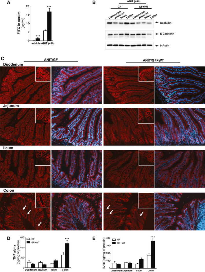
Fig. 5.

The intestinal microbiome exacerbates intestinal permeability during ANIT‐induced cholestasis. (A) Quantification of circulating FITC in serum samples from GF and GF + WT mice after vehicle and ANIT (48 hours). (B) Western blotting analysis on intestinal protein extracts from duodenum, jejunum, ileum, and colon showing reduced expression of tight junctions in GF + WT mice particularly pronounced in the colon. (C) Immunofluorescence staining on intestinal sections supporting reduced apical occludin expression in ANIT/GF + WT mice. (D) TNFα and (E) IL1β protein expression determined by ELISA on protein extracts isolated from duodenum, jejunum, ileum, and colon showing more pronounced inflammation in colons from ANIT/GF + WT mice. Values are mean ± SEM. *P < 0.05; **P < 0.01; ***P < 0.001 (vehicle/GF + WT vs. ANIT/GF + WT). Abbreviation: ELISA, enzyme‐linked immunosorbent assay.
