
Figure 3.
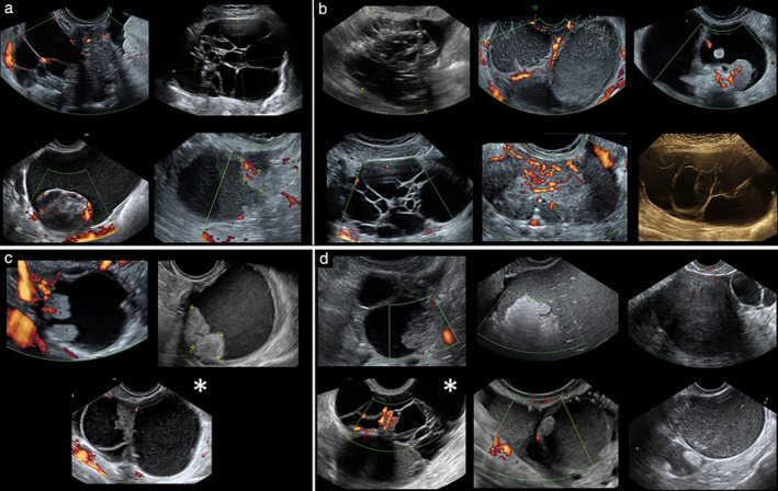
(a,b) Ten ovarian lesions that were difficult to classify by both expert subjective assessment and deep neural network model Ovry‐Dx2; final histological diagnosis was malignant or borderline in four (a) and benign in six (b). (c,d) Further nine ovarian lesions classified as inconclusive by Ovry‐Dx2; final histological diagnosis was malignant or borderline in three (c) and benign in six (d). Images marked with ( ) were also classified as inconclusive by IOTA simple rules.