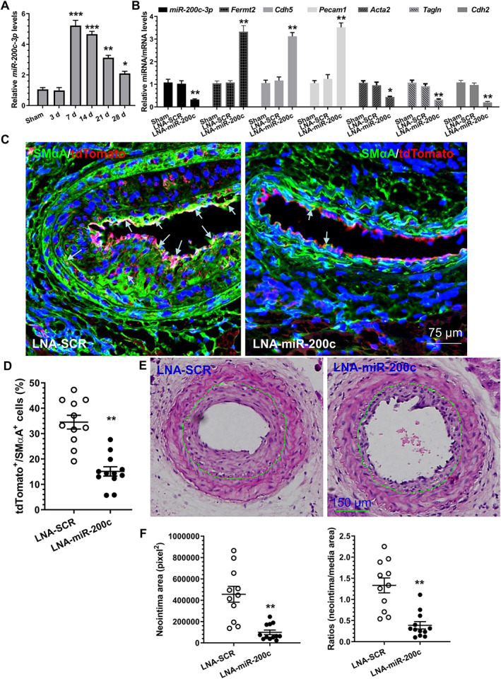
Figure 5.

miR‐200c‐3p inhibition in grafted aortas reduces EndoMT and prevents neointima formation in the grafts. (A) Increased expression of miR‐200c‐3p in grafted aortas. Total RNAs were harvested from non‐implanted (used as sham surgery) and post‐implanted aortas at the indicated times and subjected to RT‐qPCR analyses. The data presented here are mean ± SEM of five independent experiments (aortas from 3–5 mice were pooled for each experiment, n = 5 experiments). *p < 0.05, **p < 0.01, ***p < 0.001 (versus sham; one‐way ANOVA with a Tukey's post hoc test). (B) miR‐200c‐3p inhibition prevents EndoMT in the grafted aorta. Two weeks after grafting, the grafted aortas from 6–8 mice were harvested, pooled, and prepared for cell sorting. Total RNAs were extracted from tdTomato+ cells isolated from the grafted aortas treated with medium only (Sham), a scrambled negative control (LNA‐SCR), or LNA‐miR‐200c‐3p (LNA‐miR‐200c) inhibitor, respectively, and subjected to RT‐qPCR analysis. The data presented here are mean ± SEM of five independent experiments (n = 5). *p < 0.05 (one‐way ANOVA with a Tukey's post hoc test). (C, D) Decreased numbers of cells underwent EndoMT in the grafted aortas treated with LNA‐miR‐200c. Four weeks after grafting, grafted aortas were harvested and subjected to immunostaining. The data presented here are the representative images (C) and the quantitative results of tdTomato+/SMA+ (cells underwent EndoMT) (D) from 11 (LNA‐SCR) and 12 (LNA‐miR‐200c) mice, respectively. Note: arrows in C indicate cells that underwent EndoMT. (E, F) miR‐200c‐3p local inhibition decreases neointimal hyperplasia in vascular grafts. Four weeks after grafting, grafted aortas were harvested and prepared for H&E staining analyses. Representative images (E) and morphological characteristics (F) including neointimal area and neointimal/media (N/M) ratio of the implanted aortas from 11 (LNA‐SCR) and 12 (LNA‐miR‐200c) mice, respectively, are presented here. **p < 0.01 (versus LNA‐SCR, Student's t‐test).
