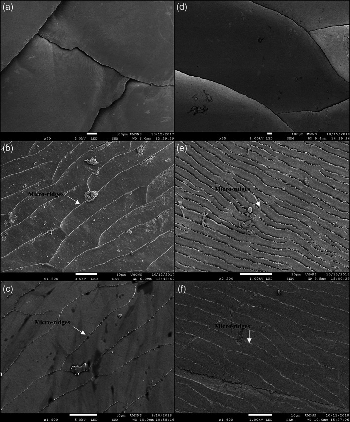
FIGURE 4.

Scanning electron micrographs of scales of Scincus scincus (left column a–c) and Eumeces schneideri (right column d–f). (a,b) Low magnification images illustrate the imbrication of the dorsal body scales of both species. Images (b and e), taken at high magnification, indicate the micro relief present on the surface of dorsal scales, whereas (c and f) focus on the microridges of the ventral scales. The white arrows indicate microridges
