Figure 1.

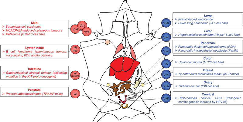
Opposite roles of murine γδ T cell subsets in cancer. Distinct γδ T cell subsets play an anti‐ (red) or pro‐ (blue) tumoural roles in various experimental mouse cancer models. Vγ1+, Vγ4+ and Vγ5+ T cell subsets producing IFN‐γ are protective in several skin cancers such as squamous cell carcinoma, 3‐methylcholanthrene (MCA)/ 7,12‐dimethylbenz]alpha]anthracene (DMBA)‐induced cutaneous tumors or B16‐F0 melanoma. γδ T cells are also overtly anti‐tumoural in: (i) spontaneous B cell lymphoma in mice lacking beta 2 microglobulin (β2m) and/ or perforin (Pfn), (ii) gastrointestinal stromal tumors due to activating mutation in the KIT proto‐oncogene, via GM‐CSF, and (iii) prostate adenocarcinoma in TRAMP mice. In contrast, Vγ6+ γδ17 T cells play pro‐tumor roles in Kras‐induced lung cancer and Lewis lung carcinoma, as well as in ID8 ovarian cancer and in Human papillomavirus (HPV)‐induced cervical squamous cell carcinoma (SCC) models. Other IL‐17‐producing γδ T cells, especially Vγ4+ T cells, are detrimental in (i) Hepa1‐6 hepatocellular carcinoma, (ii) pancreatic adenocarcinoma in PDA and pancreatic intraepithelial neoplasia (PanIN) models, and (iii) KEP breast cancer metastatic mouse model.
