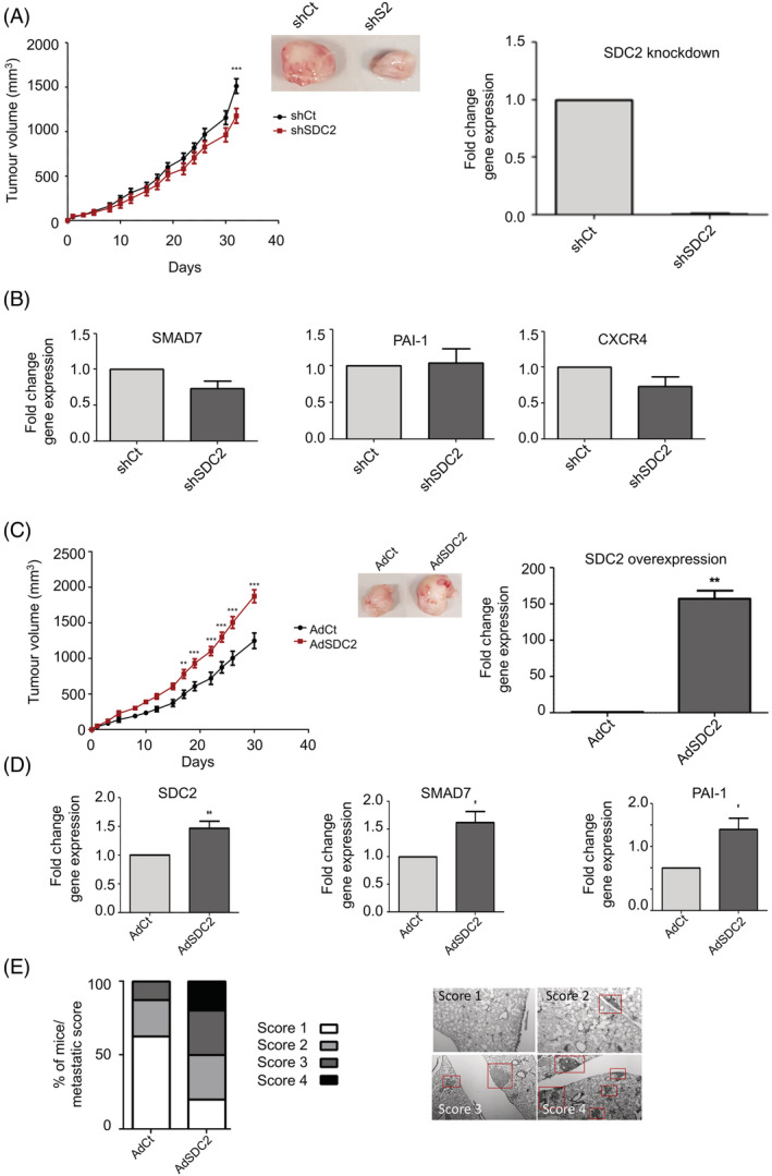
FIGURE 3.

Manipulation of SDC2 within the stromal cell compartment of xenograft tumours effects breast carcinogenesis. A, Orthotopic xenograft tumours were established by coinjecting MDA‐MB‐231 with shSDC2‐transduced TASCs or shCt‐transduced TASCs into the mammary fat pad of immune‐compromised mice at a ratio of 1:10 (TASCs:MDA). Tumours containing shSDC2‐TASCs (n = 9) showed significantly lower growth rates when compared to control shCt‐TASC tumours (n = 10). RT‐qPCR analysis showing shSDC2‐transduced TASCs have reduced SDC2 RNA levels compared to adenovirus shCt‐transduced TASCs. B, RNA was prepared from xenograft tumours and RT‐qPCR was performed to compare the expression of TGFβ‐regulated genes between shCt and shSDC2 expressing tumours (n = 5/group). C, Orthotopic xenograft tumours were established as described earlier. Tumours containing TASCs overexpressing SDC2 (AdSDC2) (n = 10) showed significantly higher growth rates when compared to control tumours (AdCt) (n = 9). RT‐qPCR analysis showing AdSDC2‐transduced TASCs have increased SDC2 RNA levels compared to adenovirus AdCt‐transduced TASCs. D, RNA was prepared from xenograft tumours and RT‐qPCR was performed to determine the effect of SDC2 modulation within TASCs upon the expression of TGFβ‐regulated genes. (n = 5/group). E, Approximately 12 weeks after TASC:MDA injection, lungs were removed and examined for metastatic nodules by H&E staining. (AdCt [n = 9], AdSDC2 [n = 10]). The bar graph indicates the metastatic score/lung from the various experimental groups. Red boxes indicate metastatic lesions. *P ≤ .05; **P ≤ .01; ***P ≤ .001 [Color figure can be viewed at wileyonlinelibrary.com]
