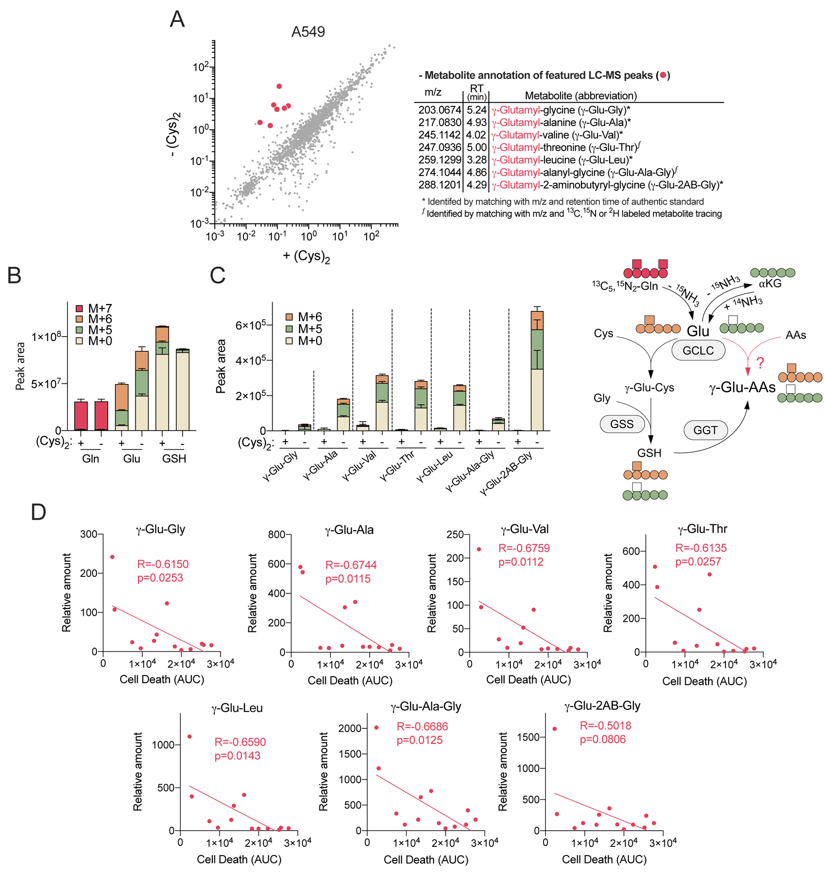
Figure 2. Cystine starvation induces glutamate-derived γ-glutamyl-peptide accumulation.

(A) Scatter plot comparison of non-targeted metabolomics features in A549 cells cultured under cystine replete (+Cys2) and starved conditions (-Cys2) for 4 hrs (left). The mean intensity of median-normalized LC-MS peaks of each group (N=3) are plotted on the axes, and each dot represents an individual LC-MS peak. The LC-MS peaks that highly accumulated under cystine starvation (red dots) were further identified and annotated (right). (B-C) A549 cell 13C5, 15N2-Gln tracing into (B) Gln, Glu, GSH, and (C) γ-Glu-peptides following culture in cystine replete or starved conditions for 4 hrs (N=3). (D) Correlation between ferroptotic cell death (AUC, from Figure 1A) and the levels of γ-Glu-peptides across 13 NSCLC cell lines. The γ-Glu-peptides were analyzed following cystine starvation for 12 hrs and normalized to the mean value of H1581 cells under cystine replete conditions (N=13). For B-D, data are shown as mean ± SD. N is number of biological replicates. For D, Pearson correlation test was used for statistical analysis.
