
Figure 4.
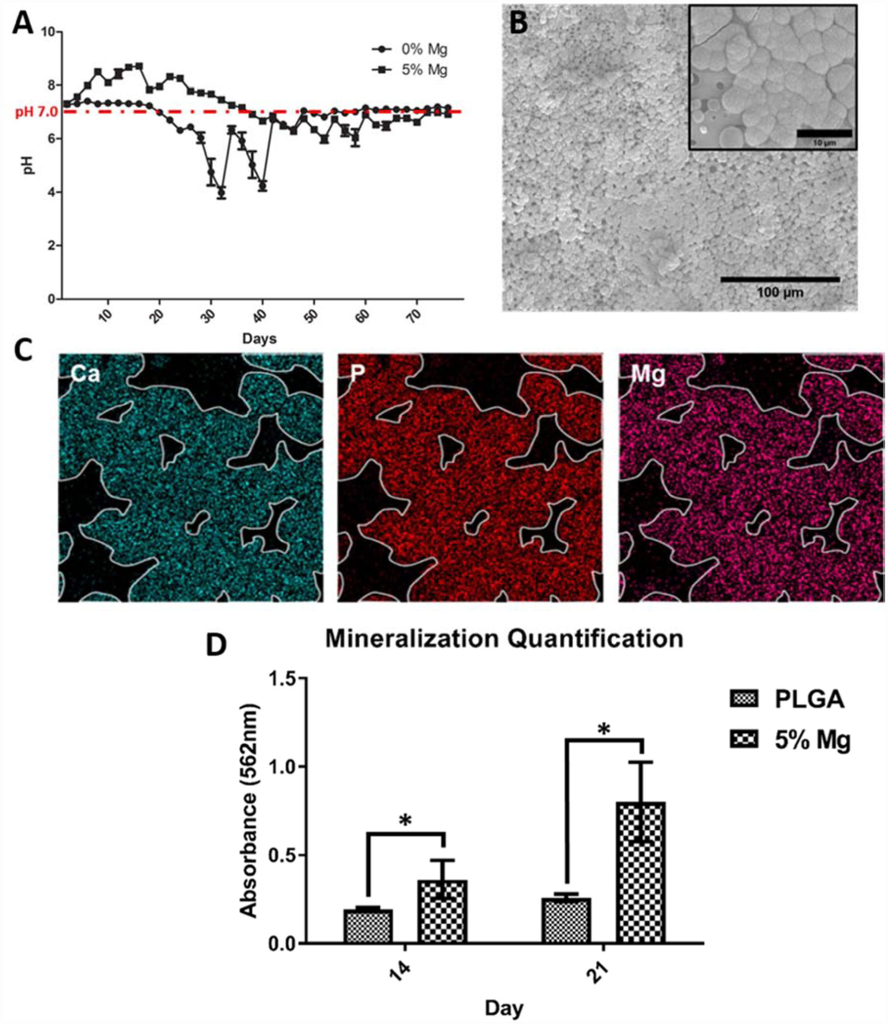
PLGA-Mg composite biomaterial neutral degradation. (A) Degradation profile of PLGA and PLGA containing 5wt% of Mg. (B) SEM image of 5% Mg:PLGA after 14 days immersed in Simulated Body Fluid (SBF), insert clearly show apatite crystal growth. (C) EDAX spectra of calcium apatite deposits on 5% Mg:PLGA after immersion in SBF (D) Alizarin red quantification of PLGA and Mg:PLGA samples after culture with MC3T3-E1 cells. Reproduced from Ref # with permission of IOP Publishing, copyright 2018 (14).