Fig. 5.
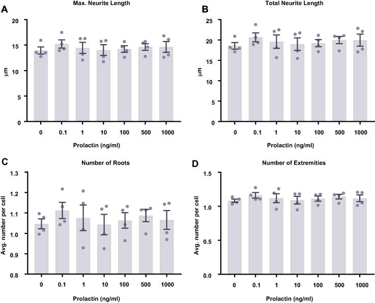
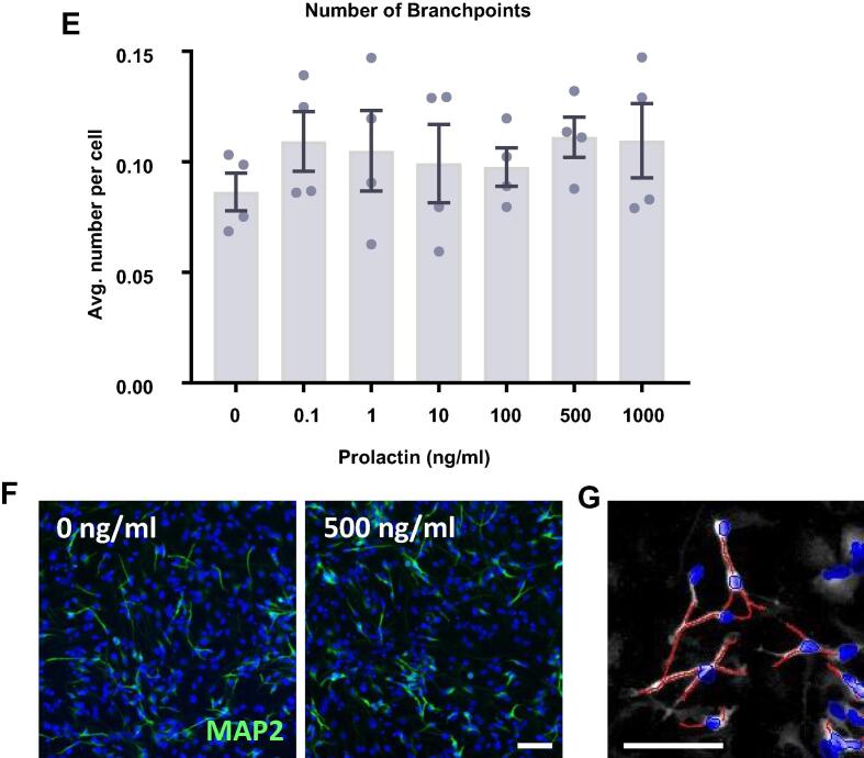
Neurite outgrowth analysis of prolactin-treated hippocampal progenitor cells after 7 days of differentiation. Following 7 d of differentiation in the presence of prolactin, cells were co-stained for MAP2 and DAPI. Columbus software was used to identify neurons based upon nuclear DAPI staining and cytoplasmic MAP2 staining. The neurites of MAP2+ neurons were traced and the number, length and branching were quantified. (A) Prolactin had no effect on the maximum neurite length present in each condition. (B) Prolactin did not impact the total length of neurites traced in each well. (C) There was no impact on the number of roots corresponding to the number of neurites originating at the cell body border, calculated as the average number per cell. (D) There was also no impact on the number of extremities corresponding to the number of ends of individual neurites, calculated as the average number per cell. (E) Prolactin also had no impact on the number of nodes or points of intersection between two or more neurite segments, calculated as the average number per cell. Group differences were detected using a one-way ANOVA. Significantly different groups were considered when p < 0.05. N = 4 for all groups. (F) Representative images of the 0 and 500 ng/ml prolactin treatment groups illustrating the similar neurite outgrowth. DAPI nuclear stain shown in blue and MAP2 shown in green. (G) Example of the automated neurite tracing performed by Columbus. Group differences were detected using a one-way ANOVA. Significantly different groups were considered when p < 0.05.


