Fig. 9.
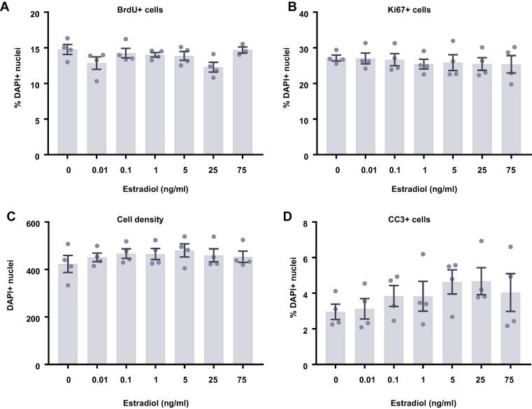
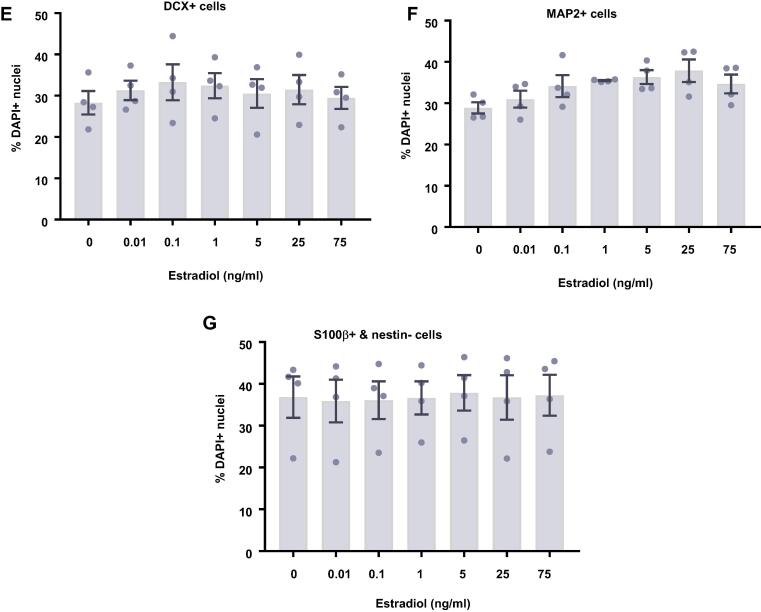
Cellular marker quantification following 7 d of differentiation with estradiol. HPCs were treated with differing concentrations of estradiol during both the 48 h proliferation phase and 7 d differentiation phases of the cellular assay. Each graph indicates the proportion of DAPI-positive cells staining positively for each marker. (A) Estradiol treatment had no impact on the proportion of cells synthesising new DNA as indicated by BrdU staining. (B) It also did not affect the number of cells in the cell cycle as indicated by KI67 staining. (C) There was also no effect of estradiol treatment on cell density. (D) There was no difference in the proportion of apoptotic cells as shown by CC3 staining. (E) Estradiol treated had no impact on the proportion of cells staining positively for DCX or (F) MAP2 indicating no change in neuronal differentiation. (G) There was also no effect of prolactin treatment on astrocytic cells which were positive for S100β but negative for nestin. Group differences were detected using a one-way ANOVA followed by Bonferroni’s post-hoc test. Significantly different groups were considered when p < 0.05. N = 4 for all groups.


