Fig. 3.

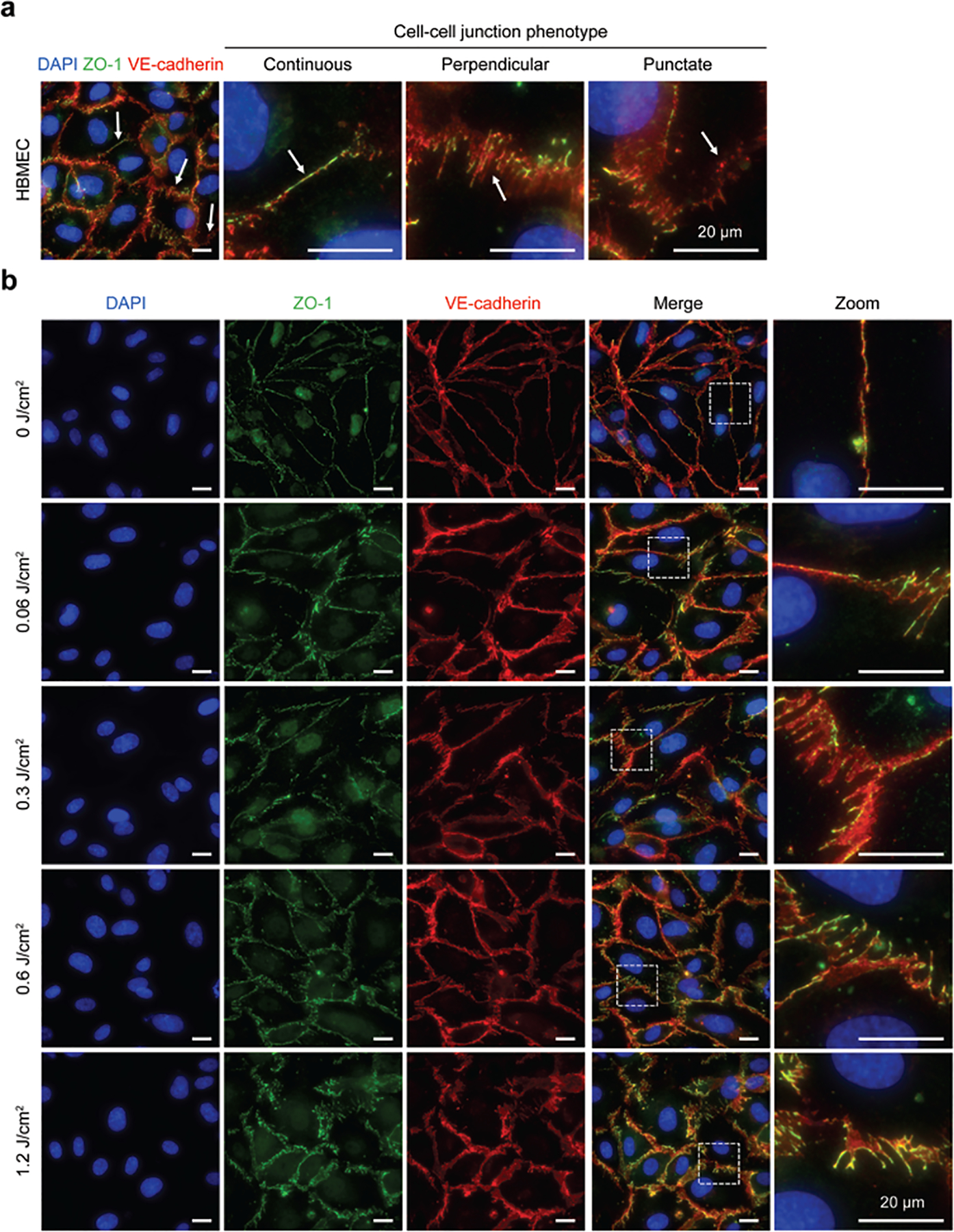
PDP modulates endothelial cell-cell junction phenotype. At 90 minutes post-priming, cells were fixed, stained, and imaged, (a) Representative fluorescence image of PDP modulated HBMEC with stained nuclei (blue), ZO-1 (green), and VE-cadherin (red). White arrows show regions of different cell-cell junction phenotypes: continuous linear junctions, perpendicular junctions, and dotted punctate junctions. (b) Representative fluorescence images of non-treated cells (0 J/cm2) and photodynamic primed cells (0.06–1.2 J/cm2). PDP generates immature HBMEC monolayers by decreasing linear continuous junctions and increasing perpendicular junctions, in a light dose-dependent manner. All scale bars are 20 μm.
