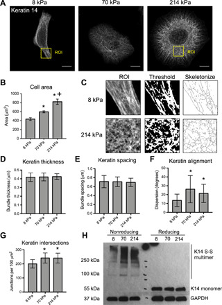
Fig. 1. Matrix rigidity regulates structural remodeling of the keratin cytoskeleton.

(A) Representative immunofluorescence images of K14 in HaCaT keratinocytes cultured for 24 hours on PA gels with elastic moduli of 8, 70, and 214 kPa. Images were acquired using Zeiss Airyscan confocal microscopy. Scale bars, 10 μm. (B) Quantification of average cell area on PA hydrogels. Data represent means ± SEM of N = 3 experiments (40 to 50 cells per experiment). *P < 0.05 compared to 8 kPa. +P < 0.05 compared to 70 kPa. (C) Schematic of the ImageJ analysis protocol for K14 bundles, including selection of a region of interest [ROI; labeled in (A)], thresholding, and skeletonization. (D) Quantification of average K14 bundle thickness and (E) spacing was performed on ROIs within the cytoplasm using the BoneJ plug-in. (F) Keratin alignment was quantified by the mean dispersion (SD) of K14 bundle angles within the whole cell using the Directionality plug-in. (G) Keratin bundle intersection was estimated by quantifying the mean density of junctions within skeletonized images of K14 using the Skeleton plug-in. All keratin data represent means ± SD of N = 30 cells from three experiments. *P < 0.05 compared to 8 kPa. (H) Western blot analysis of keratin cross-linking for cell lysates prepared with (reducing) or without (nonreducing) β-mercaptoethanol and probed for K14 and GAPDH.
