


Fig. 2. Evidences for oligoamidine 3’s two distinct antimicrobial mechanisms of action, membrane disruption, and DNA binding.
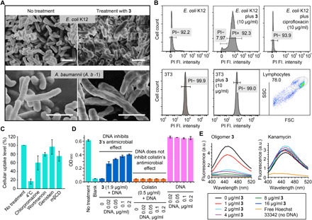
(A) 3-treated E. coli (K12) (top) and MDR A. b -1 (bottom) cells showing clear evidence of membrane damage, exhibiting fragments and wrinkled surfaces, similar to previous reports on surfactant-based antimicrobial polymers. Scale bars, 4 μm. (B) Results of membrane permeability assay using E. coli, 3T3 cells and propidium iodide (PI), showing bacterial cell membrane disruption by the addition of 3. PI is a fluorescent dye that can only penetrate compromised cell membranes, so its uptake into cells indicated membrane disruption. Compound treatment time was 4 hours for E. coli. The membrane of the NIH/3T3 cells were not affected by 3 even over an extended time period (24 hours). Fl., fluorescence. (C) Exploration of 3’s cellular internalization mechanism. Chlorpromazine, wortmannin, mβCD/genistein, and 4°C condition, respectively, inhibit clathrin-mediated endocytosis, macropinocytosis, caveolae-mediated endocytosis, and energy-dependent endocytosis in general. (D) Inhibition of 3’s activity by DNA. Externally added DNA substantially inhibited oligomer 3’s antimicrobial effect against MDR A. b -1, whereas colistin’s efficacy was minimally affected by added DNA. (E) Fluorophotometric studies showing that 2 μg/ml of 3 was able to displace >80% of the minor groove binding dye, Hoechst 33342 (4 μg/ml), bound to double-stranded DNA (dsDNA), while kanamycin was not able to incur any change in Hoechst’s fluorescence. a.u., arbitrary units.