
Fig. 1. A reporter-based CRISPR screen identifies KLF11 as a negative regulator of osteosarcoma CSCs.
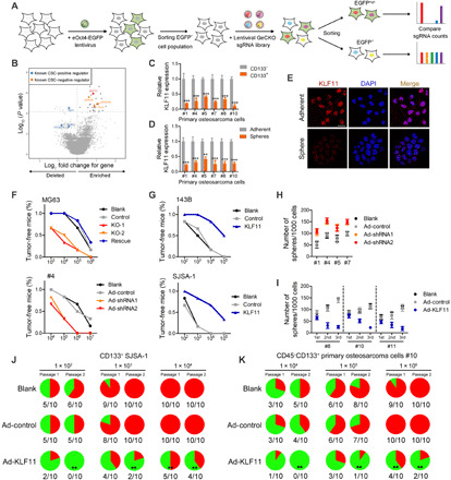
(A) Experimental design of an eOct4-EGFP reporter–based genome-wide CRISPR screen. (B) Volcano plot displaying the log2 fold change and adjusted P value for all sgRNAs identified in the screen. Negative regulators with a threshold of FDR < 0.05 are labeled as small dark gray dots, selected genes with known function as large blue and orange dots, and KLF11 as large red dot. (C) qRT-PCR analysis of KLF11 expression in CD133+ and CD133− cells from primary osteosarcoma cells (n = 3). (D) qRT-PCR analysis of KLF11 expression in spheres and adherent cells from primary osteosarcoma cells (n = 3). (E) Immunofluorescence detection of KLF11 (red) in primary osteosarcoma #4 spheres and adherent cells. The nuclei were stained with 4,6-diamidino-2-phenylindole (DAPI) (blue). Scale bar, 25 μm. (F) In vivo limiting dilution assay of KLF11-deleted and ectopic KLF11 rescue MG63 cells, and KLF11-knockdown primary osteosarcoma cells (sample #4). KO, knockout. n = 6 for each group. (G) In vivo limiting dilution assay of KLF11-overexpressing and control osteosarcoma spheres (143B and SJSA-1). n = 6 for each group. (H) Sphere formation assay of KLF11-knockdown primary osteosarcoma cell. (I) Sphere formation assay of KLF11-overexpressing primary osteosarcoma cells (n = 3). (J) Incidences of tumorigenesis of CD133+ SJSA-1 cells infected with indicated adenovirus in serial transplantation models. **P < 0.01 compared with untreated CD133+ SJSA-1 cells in the first inoculation by Fisher’s exact test. (K) Incidences of tumorigenesis of CD45−CD133+ primary osteosarcoma cells (sample #10) infected with indicated adenovirus in serial transplantation models. **P < 0.01 compared with untreated primary osteosarcoma cells in the first inoculation by Fisher’s exact test.