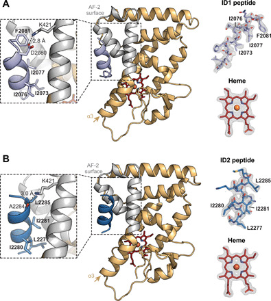
Fig. 2. Crystal structures of REV-ERBβ LBD cobound to heme and NCoR ID peptides.

(A) Structure of REV-ERBβ LBD (light orange cartoon with the AF-2 surface in gray) cobound to heme (red sticks) and NCoR ID1 peptide (light purple cartoon) (PDB 6WMQ). (B) Structure of REV-ERBβ LBD (light orange cartoon with the AF-2 surface in gray) cobound to heme (red sticks) and NCoR ID2 peptide (dark blue cartoon) (PDB 6WMS). Insets to the left of the structures highlight the ID peptide CoRNR box motif residues (in bold) and conserved charge clamp interaction with K421. Omit maps (2Fo − Fc, contoured at 1σ) for the ID peptides and heme are shown to the right of the structures with CoRNR box motif residues indicated.
