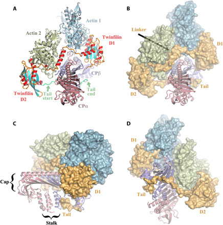
Fig. 1. The twinfilin/CP/actin complex.

(A) Front view of the pentameric complex in cartoon representation. CP consists of two subunits, α-subunit (CPα) and β-subunit (CPβ). Twinfilin comprises two ADF-H domains (D1 and D2), a linker between D1 and D2 that includes a helix (residues 151 to 165), and a C-terminal tail (Tail; residues 316 to 342, the last eight amino acids are disordered). Actin subunit 1 is bound to twinfilin D1, and subunit 2 to twinfilin D2. The twinfilin secondary structure elements are colored differentially, helices in red, strands in cyan, and loops and extended regions in orange, with the Tail in lime green. (B) Front view [same as in (A)], in which the actin subunits and twinfilin are represented as surfaces. Twinfilin is shown entirely in orange. (C and D) Back and side views, respectively.
