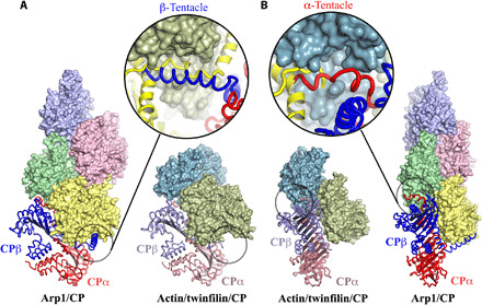
Fig. 4. Comparison of the CP-binding mode to Arp1 in the dynactin complex (Arp1/CP) with the binding mode to actin in actin/twinfilin/CP complex.

(A and B) Focus on the β- and α-tentacles, respectively, which are highlighted by black circles. The β-tentacle is disordered and unbound in the actin/twinfilin/CP complex, and the α-tentacle is partially bound and ordered, relative to the dynactin complex. Enlargements of the tentacle regions show the superimpositions of the CP (α- and β-subunits colored red and blue, respectively) from the dynactin complex on to actin (green and teal) and twinfilin (yellow) from the actin/twinfilin/CP complex. (A) Enlargement, the twinfilin linker (yellow) binds to the β-tentacle–binding site on actin 2 (green). (B) Enlargement, the N terminus of twinfilin D1 (yellow) occupies half of the α-tentacle–binding site on actin 1 (teal).
