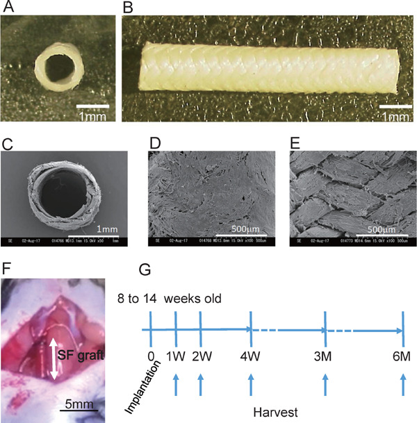
Fig. 1.

Preparation of silk fibroin graft and implantation into mouse carotid artery
A and B, Silk fibroin-based graft (0.9 mm in diameter). Cross-sectional image (A) and whole image (B) (scale bar, 1 mm). C to E, Scanning electron microscope (SEM) images of graft. Cross-sectional images (C), outside of graft (D) and inside of graft (E) (scale bar, 1 mm in C, 500 µm in D and E). F, The graft was implanted into the right carotid artery of a mouse with a cuff technique (SF, silk fibroin. scale bar, 5 mm). G, Study protocol of graft implantation. Grafts were implanted into 8- to 14-week-old male C57BL/6 mice and harvested at 1, 2, and 4 weeks and 3 and 6 months.
