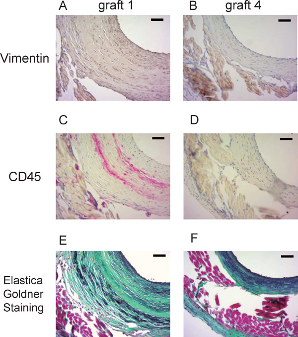
Fig. 6.

Types of cells which contribute to neointimal formation
The sections near the midline of the patent grafts harvested at 6 months with the largest ratio of neointimal area to outline area (graft 1) and the smallest ratio of neointimal area to outline area (graft 4) were stained with anti-vimentin antibody (A and B) and anti-CD45 antibody (C and D). Collagen and elastin fibers were stained by Elastica–Goldner staining (E and F). Green area indicates collagen fibers, and purple area indicates elastin fibers (scale bar, 50 µm).
