
Author response image 1. (A) Percentage (%) and (B) number (#) of total peritoneal DCs (live Lin- CD11c+ MHC II+) and MCMV-infected DCs (m04+ cells from total DCs).
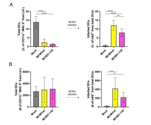
BALB/c mice were i.p. infected with 106 PFU/mice of MCMVwt and MCMVΔm138. DCs were isolated from the peritoneal cavity 6 hr post-infection and analyzed by flow cytometry. Results represent two merged individual experiments (mock n=8, MCMVwt n=6, MCMVΔ138 n=7). One-way ANOVA test was used for statistical analysis. Graphs show mean with SEM as error bars. ****, p ≤0.0001; ***, p ≤ 0.001; **, p ≤ 0.01; *, p ≤ 0.05.