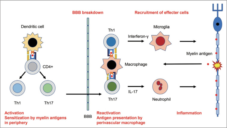
Figure 2.

Possible autoimmune mechanisms underlying MS. Naive CD4+ T cells are first sensitized by myelin antigens in the peripheral lymph nodes and differentiate into myelin antigen-specific Th1 or Th17 cells in MS. These peripherally activated Th1 or Th17 cells express increased amounts of adhesion molecules that allow them to pass through the BBB. The Th1 and Th17 cells are reactivated by perivascular macrophages, leading to the invasions into the CNS parenchyma. Reactivated Th1 and Th17 cells secrete interferon-γ and IL-17, which induces the infiltration of activated microglia and neutrophils, respectively. These effector cells secrete downstream proinflammatory or cytotoxic cytokines/chemokines, leading to demyelination or neuronal damages. These autoimmune processes are classified into several steps: activation in the periphery, BBB breakdown, reactivation and inflammation. It is uncertain whether similar autoimmune mechanisms underlie immune-mediated cerebellar ataxias. MS: multiple sclerosis, BBB: blood-brain barrier, CNS: central nervous system.
