. 2020 Nov 17;17(1):1–10. doi: 10.3988/jcn.2021.17.1.1

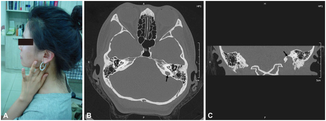
Fig. 1. A case of a high jugular bulb. (A) A 33-year-old female presented at our clinic with pulsatile tinnitus on her left ear with a 2-year history. The tinnitus was diminished by pressing just posterior to the left mandibular angle with her two fingers. There were no significant findings in blood tests for anemia, thyroid dysfunction, and hematological malignancy. Axial (B) and coronal (C) images of temporal-bone computed tomography revealed jugular-bulb diverticulum (arrows) at the level of the oval window without bony dehiscence, suggesting a high jugular bulb on the left side. Ten years had passed since her first visit to our clinic. In a recent follow-up call on June 25, 2020, the patient reported that her tinnitus was continuing but that it did not disturb her daily life or sleep. Written consent was obtained from the patient.

Fig. 1