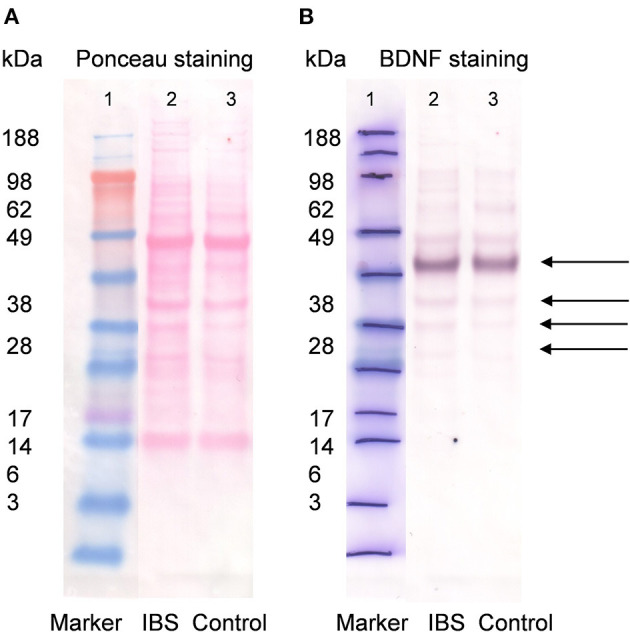
Figure 1.

Western blot for BDNF (1:5,000) in IBS patients and control subjects. Lane 1 contains the molecular weight standards. Lane 2 contains colonic wall protein obtained by deep tissue biopsy from mucosa/submucosa in patients with IBS (14 female and six male, pooled sample) and lane 3 colonic wall protein from control subjects (two female and eight male, pooled sample, A). Same blot after washout of ponceau staining solution and application of primary anti-BDNF antibody (1:5,000) and secondary antibody goat anti-rabbit AP (1:2,000). Lane 1 contains molecular standards highlighted with pen. Lane 2 and 3 show detection of multiple bands with similar intensity for patients with IBS and healthy controls, respectively. For BDNF (UniProt P23560) multiple Western Blot bands are possible and expected. The strongest band was found between 49 and 62 kDa most likely representing glycosylated prepro-/pro BDNF dimer. Weaker bands were detected at ~ 45, 38, and ~ 30 kDa likely related to prepro-/pro BDNF (B).
