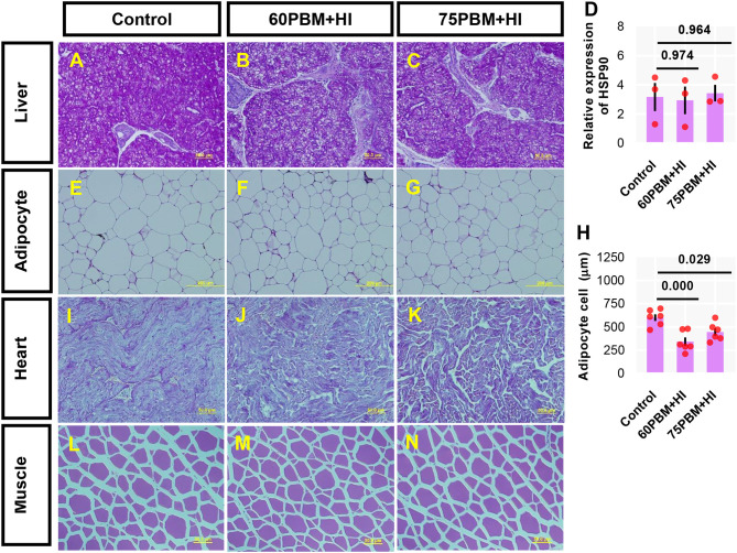
Figure 2.
Light microscopy of the liver (A–C) [periodic acid–Schiff (PAS) stain, ×40 magnification), adipose cell size (E–G) (PAS stain, ×20 magnification), heart (I–K) (PAS stain, ×40 magnification), and muscle (L–N) (PAS stain, ×40 magnification) of juvenile barramundi 6 weeks post-feeding with control, 60% FM replacement diet (60PBM + HI) and 75% FM replacement diet (75PBM + HI), supplemented with 10% full-fat HI larvae. (D,H) Variation in the expression of hepatic HSP90 (n = 3) and adipocyte cell size (n = 6) in response to different levels of PBM supplemented with HI. The results represent mean ± SEM. P-values on the top of the bar with scatter dot plot denote significant differences between control vs. 60PBM + HI-fed and 75PBM + HI-fed fish (ordinary one-way ANOVA, followed by Dunnet's multiple comparison test, P < 0.05).

