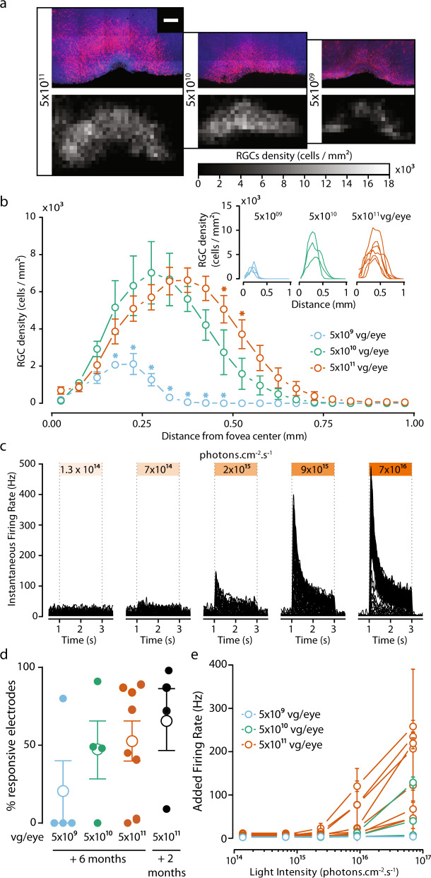
Fig. 2. AAV2.7m8–ChR-tdT induces high-density long-term expression in the perifoveal area in non-human primates.
a, top: Projections of confocal stack stitches showing perifoveal areas of retinas treated 6 months earlier with three different doses of the vector (5 × 10¹¹, 5 × 10¹⁰, and 5 × 10⁹ vg/eye, respectively left to right). ChR-tdT-expressing cells are shown in red, whereas DAPI staining of the nuclei is shown in blue. Scale bar: 200 µm. Bottom: Density maps of ChR-tdT-positive RGCs for the three hemifoveas showed on top. b Density profiles of ChR-tdT-expressing RGCs relative to retinal eccentricity for the three vector doses tested. Density is expressed as the mean value for all retinas analyzed (P < 0.05, two-way ANOVA, multiple comparisons). The individual retina profile is shown in the inset (n = 4 for 5 × 10⁹, n = 3 for 5 × 10¹⁰, and n = 6 for 5 × 10¹¹ vg/eye). c Spike density function for all responsive electrodes of a retina, treated with 5 × 10¹¹ vg, in response to different light levels. d Responsive electrode fraction, measured in MEA experiments, for the three doses after 6 months of expression, compared to 2 months of expression for 5 × 10¹¹ vg.eye (same data as Fig. 1); no significant differences shown (Wilcoxon–Mann–Whitney test). Filled circle: value for an individual retina; open circle: mean for all the retinas ± SEM. e Mean±SEM additional firing rate per responsive retina for the three doses, at different light levels, at 590 nm ±15 nm, see Supplementary Table S2 for the statistical analysis of the dose-dependent response.

