Figure 1.
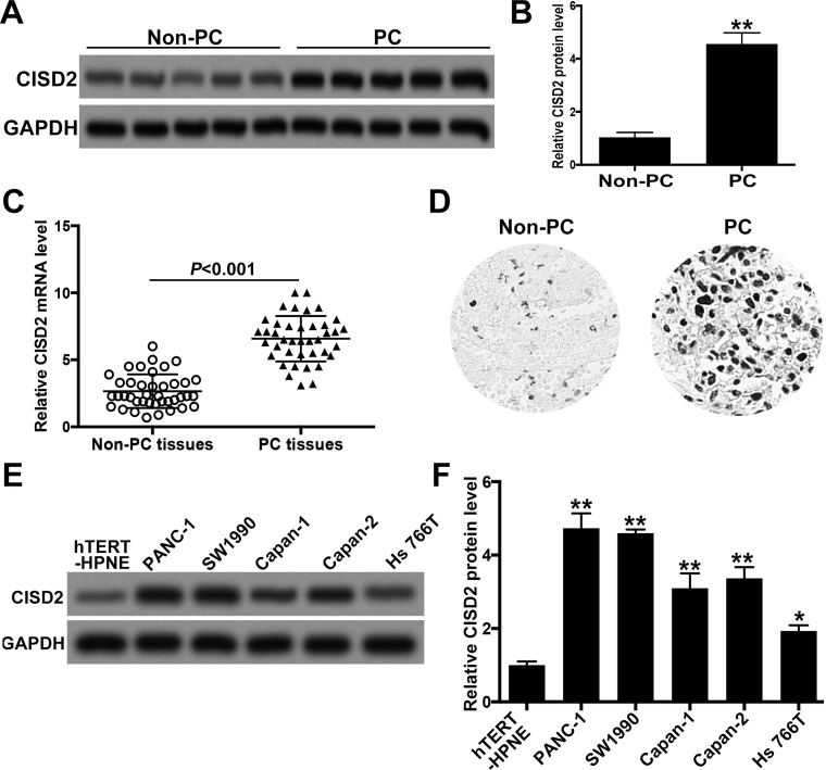
CISD2 was increased in pancreatic cancer samples and cell lines. (A) We collected 40 fresh samples of pancreatic cancer (PC) and their adjacent non-pancreatic cancer (non-PC) tissues, and Western blot was used to determine the level of CISD2. (B) Relative level of CISD2 in (A) was measured using ImageJ and normalized to GAPDH. (C) qRT-PCR was applied to test the level of CISD2 mRNA in the collected PC and non-PC tissues. (D) Immunohistochemistry staining was used to confirm the increase in CISD2 level of PC tissues compared with non-PC tissues. (E) Western blot was applied to determine the level of CISD2 in several pancreatic cancer cell lines (PANC-1, SW1990, Capan-1, Capan-2, and Hs766T). hTERT-HPNE is an immortalized normal pancreatic cell line and acts as a control. (F) Relative level of CISD2 in (E) was measured with ImageJ and normalized to GAPDH. Data were presented as mean ± SD from at least three independent experiments. *p < 0.05 and **p < 0.01 compared with control group.

