
Figure 1.
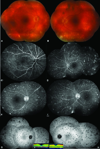
Composite of color fundus showing 360° scattered yellowish–gray spot like aggregates, (A) right eye, (B) left eye. Early venous phase of composite fluorescein angiographic picture exhibiting staining of these spot-like lesions, (C) right eye, (D) left eye. Late venous phase of composite fluorescein angiographic picture showing leakage from the optic disc, (E) (right eye), (F) left eye. Mid-phase of composite indocyanine green picture demonstrating the hypocyanescent widespread spot-like opacities, (G) right eye, (H) left eye. Normal foveal contour on optical coherence tomography, (I) right eye, (J) left eye.