Figure 3.

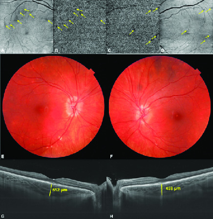
(A&B) Choriocapillaris slab of montage optical coherence tomography angiography and en-face OCT image of the right. (C&D) Left eyes revealing the patchy ischemia of choriocapillaris (arrows). Color fundus pictures taken at the last visit showing slightly blurred disc margins with less apparent old scars with no new lesion. (E) Right eye and (F) left eye. (G) Swept source optical coherence tomographic subfoveal choroidal thickness was 512 μm in the right eye and (H) 498 μm in the left eye.
