Figure 2.

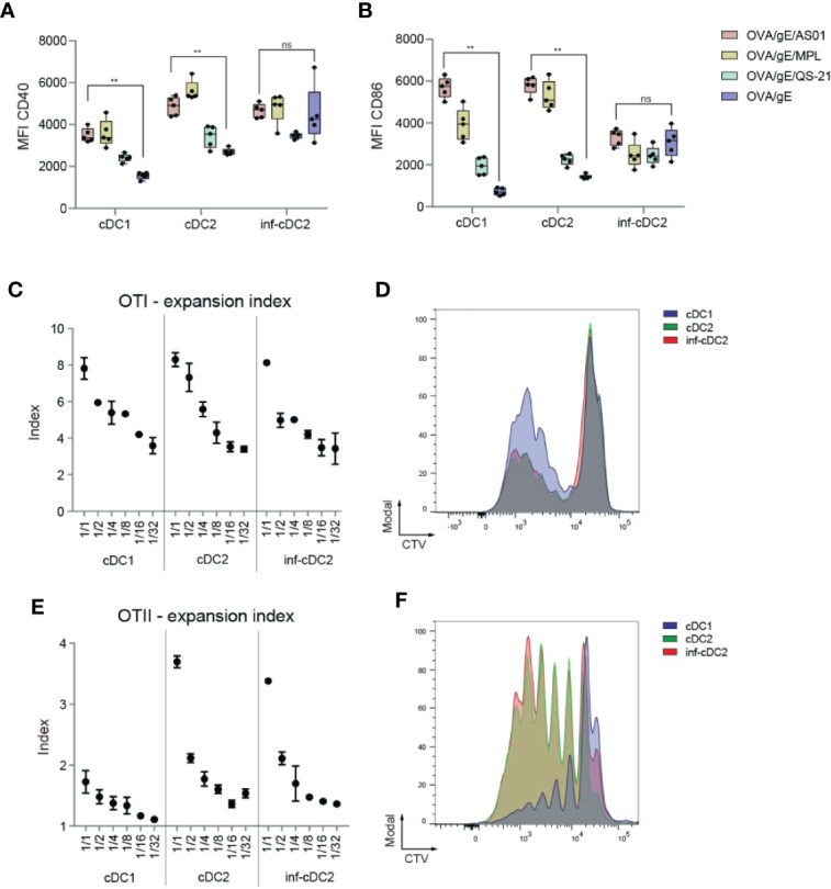
AS01 activated cDCs effectively prime antigen-specific T cells. (A, B) Expression of CD40 (A) and CD86 (B) shown as MFIs by cDC subsets in the dLN 24 h after i.m. (M. gastrocnemius) immunization with OVA/gE antigen (0.5/2.5 µg) formulated in 2.5 µg AS01, MPL, QS-21, or buffer per injection site. AS01, MPL, and QS-21 are all in liposome. Data analyzed with Two-way ANOVA and Tukey’s multiple comparisons test. Data are representative of at least two independent experiments (n = 5 mice per group). **p < 0.01, ns, non-significant. (C) Expansion index of CD8+ OVA-specific TCR transgenic T cells (OTI) cocultured for 3 days with the different migratory cDC subsets sorted from pooled dLNs (n = 80 mice) 24 h after i.m. immunization with OVA/AS01 (5/1 µg) per injection site. (D) Proliferation prolife of CTV-labeled OTI T cells cocultured in 1:4 DC:T cell ratio with the different migratory cDC subsets for 3 days sorted from pooled dLNs (n = 80 mice) 24 h after i.m. immunization with OVA/AS01 (5/1 µg) per injection site. (E) Expansion index of CD4+ OVA-specific TCR transgenic T cells (OTII) cocultured for 4 days with the different migratory cDC subsets sorted from pooled dLNs (n = 80 mice) 24 h after i.m. immunization with OVA/AS01. (5/1 µg) per injection site (F) Proliferation prolife of CTV-labeled OTII T cells cocultured in 1:1 DC:T cell ratio with the different migratory cDC subsets for 4 days sorted from pooled dLNs (n = 80 mice) 24 h after i.m. immunization with OVA/AS01 (5/1 µg) per injection site.
