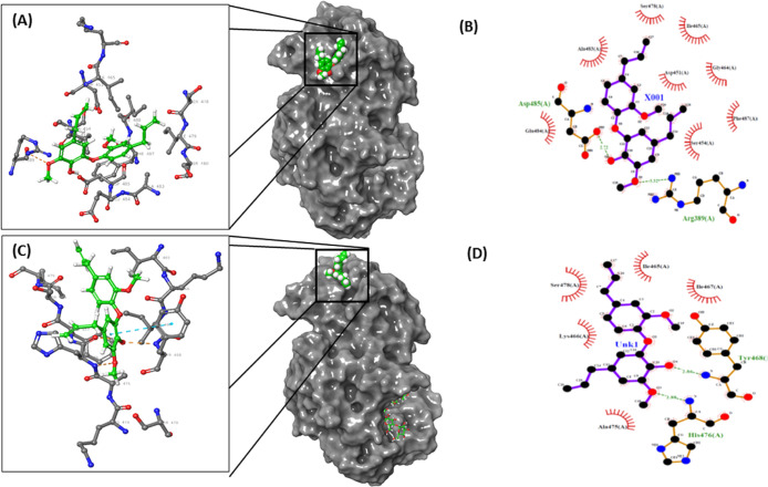
Figure 4.
Molecular docking of DDEB with HPA at Site 4. (A) and (C) Full views and close-up views of HPA–DDEB binary and HPA–maltohexaose–DDEB ternary complexes, respectively, showing H-bonds (beige dotted lines) and pi–pi interactions (cyan dotted line). In full view, DDEB is shown with CPK rendering and maltohexaose (in C) is shown as ball-and-stick rendering with carbons (green), oxygens (red), and hydrogens (white). HPA is rendered as solvent-accessible surface. The interactions of HPA and DDEB in binary (B) and ternary (D) complexes identified using LigPlot with H-bonds shown as green dotted lines and hydrophobic contacts shown as spoked arcs.

