Fig. 1.
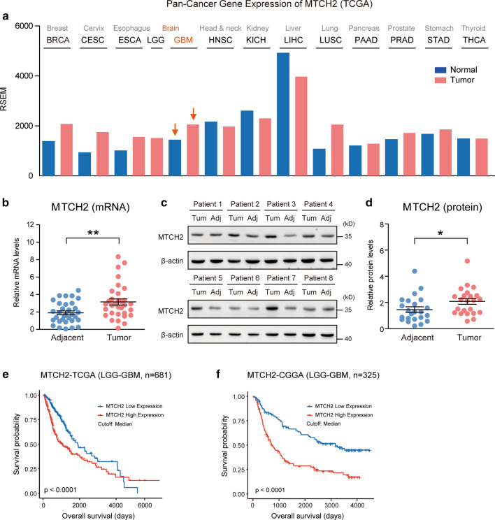
MTCH2 expression is increased in glioma tissues and indicates poor prognosis. a A Pan-cancer diagram showing the increased MTCH2 expression in brain gliomas. bReal-time PCR results showing increased mRNA level of MTCH2 in glioma tissues compared to paired adjacent brain tissues (n = 32). c, d Western blots (c) and quantifications (d) showing increased proteins of MTCH2 in glioma tissues compared to paired adjacent brain tissues (n = 23). e, f Survival analysis using clinical information from TCGA (e) and CGGA (f) dataset. Patients are divided into low and high MTCH2 groups by median expression level

