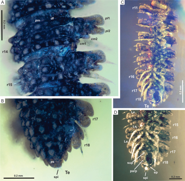
Figure 4. Myrmecodesmus antiquus sp. nov.
(A) CPAL.117, dorsal view of rings 14 and 15, showing paramedian and dorsolateral tubercles, and paranota with two caudomarginal and two paranotal lobes. (B) CPAL.117, dorsolateral view of posterior end, showing rings 16–18, and telson. (C) CPAL.117, ventral view of midbody rings and posterior end, showing rings 11–18, and telson. (D) CPAL.117, closer view at the posterior end, ventral view, showing subanal plate, paraprocts, epiproct, and spinnerets. Abbreviations: cm, caudomarginal lobe; dl, dorsolateral tubercle; ep, epiproct; Le, leg; parp, paraprocts; pl, paranotal lobe; pm, paramedian tubercle; ps, preanal sclerite; r, body ring; spi, spinneret; sup, subanal plate; Te, telson.

