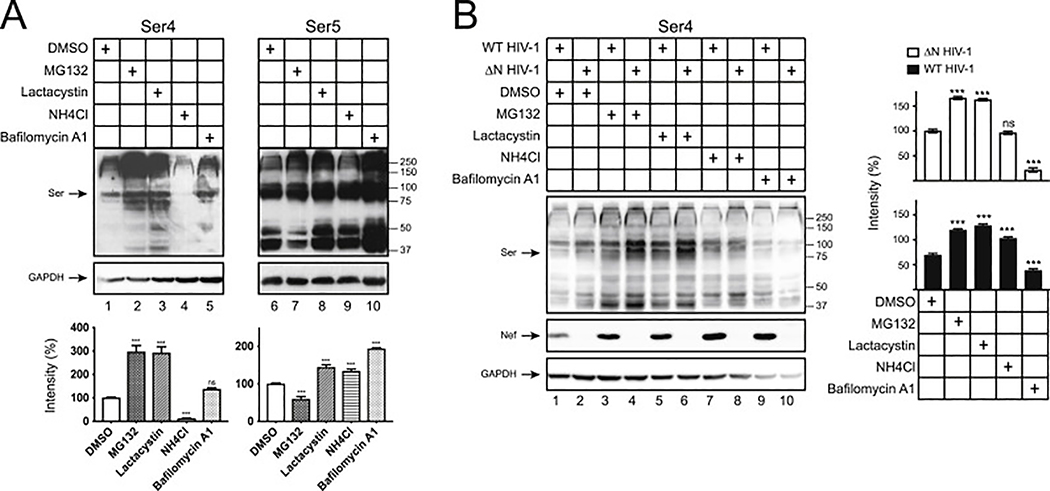
Fig. 2. Human Ser4 is degraded in proteasomes.
(A) 293T cells were transfected with 2 μg pCMV6-Ser4 or 0.5 μg pCMV6-Ser5. After treatments with DMSO (control), MG132 (10 μM), lactacystin (10 μM), NH4Cl (20 μM), or bafilomycin A1 (100 nM) for 12 h, Ser4 and Ser5 expression were determined by WB using anti-FLAG. The Ser4 or Ser5 expression levels were quantified using ImageJ and are presented as relative values. The levels of Ser4 and Ser5 samples treated with DMSO were set as 100%.
(B) 293T cells were transfected with 1 μg pH22 or pH22ΔN that produces WT or ΔN NL4–3 viruses in the presence of 1 μg pCMV6-Ser4. After similar treatments as in (A), Ser4 and Ser5 expression were determined and quantified similarly as in (A). The levels of Ser4 in the presence of ΔN HIV-1 treated with DMSO were set as 100%. Error bars represent SDs from three independent experiments. *p < 0.05, **p < 0.01, ***p < 0.001, ns, not significant (p > 0.05).

