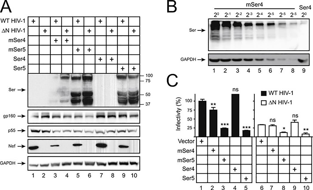
Fig. 3. Analysis of mSer4 expression and anti-HIV-1 activity.
(A) WT and ΔN HIV-1 NL4–3 viruses were produced from 293T cells in the presence of 100 ng pCMV6 vectors expressing human and murine Ser4 or Ser5. Cellular Ser4 and Ser5 expression were determined by anti-FLAG and cellular HIV-1 protein expression was detected by indicated antibodies via WB.
(B) 293T cells were transfected with 1 μg pCMV6 vectors expressing mSer4 and human Ser4. mSer4-expressing cell lysate was serially diluted and compared to human Ser4 by WB using anti-FLAG.
(C) Virions were collected from culture supernatants in (A), and viral infectivity was determined in TZM-bI cells. Infectivity is shown as relative values, with the infectivity of WT viruses produced in the presence of a control vector set as 100%. Error bars represent SDs from three independent experiments. *p < 0.05, **p < 0.01, ***p < 0.001, ns, not significant (p > 0.05).

