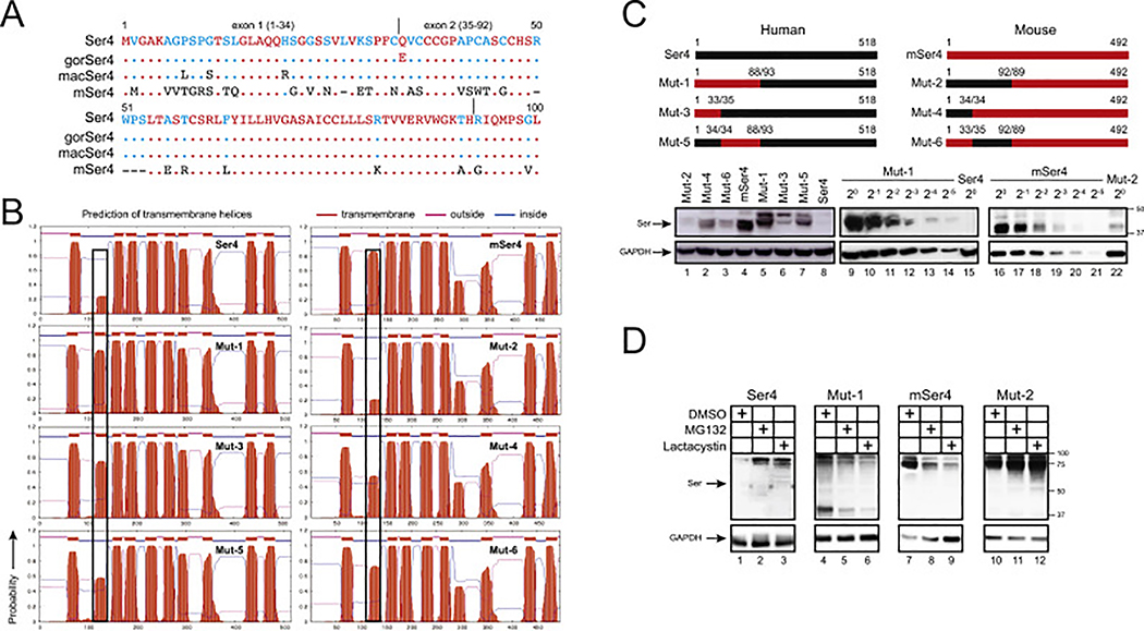
Fig. 4. Analysis of human-mouse chimeric Ser4 expression.
(A) The N-terminal amino acid sequences of human, gorilla (gor), macaque (mac), and murine (m) Ser4 proteins are aligned. Red, blue, black, and dash indicate conserved, partially conserved, non-conserved, or non-existing residues, respectively. Dots indicate identical residues. The exon 1 and exon 2 region are indicated.
(B) Membrane topology of WT and chimeric Ser4 proteins were predicted from the public TMHMM server and are presented. The putative 2nd TM domains are squared.
(C) Chimeric Ser4 mutants (Mut-1, 2, 3, 4, 5, 6) were created by swapping the indicated human Ser4 (black) and murine Ser4 (red) region and expressed from the pCMV6 vector that expresses a C-terminal FLAG tag. 293T cells were transfected with 1 μg WT or these mutant expression vectors, and their expression was compared by WB using anti-FLAG. In addition, Mut-1 and mSer4-expressing cell lysate were serially diluted and compared to human Ser4 or Mut-2 by WB using anti-FLAG.
(D) 293T cells were transfected with 1 μg pCMV6 vectors expressing human Ser4, mSer4, Mut-1, and Mut-2, and treated with DMSO (control), MG132 (10 μM), or lactacystin (10 μM) for 12 h. Their expression was determined by WB using anti-FLAG.

