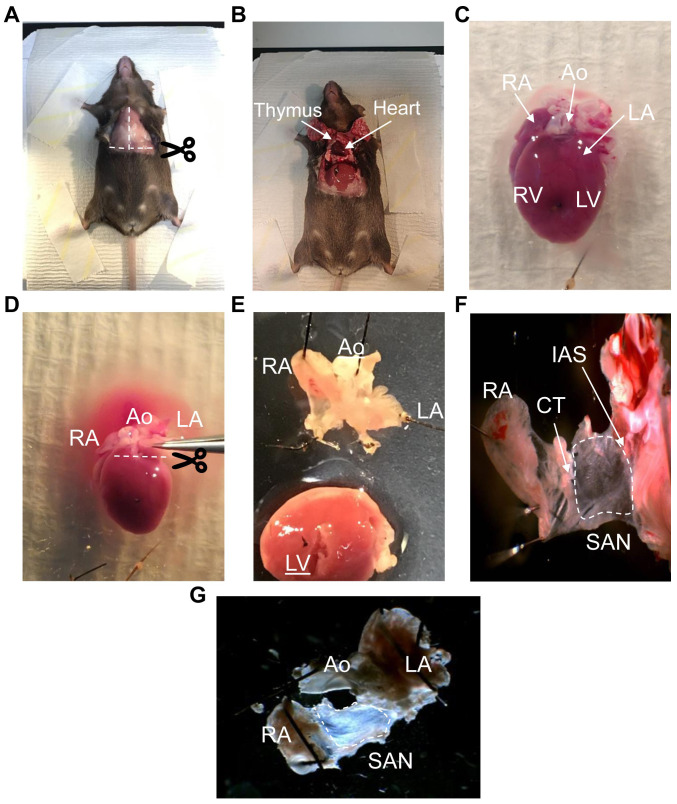
Figure 2. Dissection of the mouse heart and isolation of the SAN.
A. The anesthetized mouse is placed in the supine position and incisions made where indicated (dotted lines) to access the cardiac cavity. B. Removal of the rib cage shows the position of the heart. The heart is dissected by cutting at the aortic arch. The lungs can be removed before or after excision. C. Excised heart showing the right and left atrial appendage (RA and LA), right and left ventricles (RV and LV) and the aorta (Ao). D. The heart is pinned to the dissection dish- the dotted line shows where to cut to separate the atrial/nodal tissue from the ventricles without damaging the SAN. E. Intact SAN, RA and LA after separation from the ventricles. The incision seen on the left ventricle was used to flush out the blood from the heart before separation of the upper chambers from the ventricles. F and G. The SAN (within dotted area) and surrounding tissue after the fat and connective tissue have been removed. CT, cristae terminalis. IAS, intra-atrial septum.

