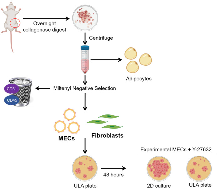
Figure 1. Isolation of rat mammary epithelial cells and fibroblasts.
The fourth mammary fat pads from female rats are removed and digested overnight. The following morning, adipocytes and red blood cells are depleted by centrifugation and lysis, respectively. The remaining cells are subjected to negative selection using Miltenyi CD31 and CD45 biotin conjugated antibodies. Next, addition of Miltenyi anti-biotin microbeads allows capture of the antibody tagged lineage positive populations on an LS column with a Miltenyi QuadroMACS magnet. The eluted MECs and fibroblasts are cultured in ultra-low attachment (ULA) plates for 48 hours to deplete fibroblasts. Purified MECs are then maintained in ULA with the addition of Y-27632 or plated into 2D culture for experimental analysis. Figure 1 was created with BioRender.com.

