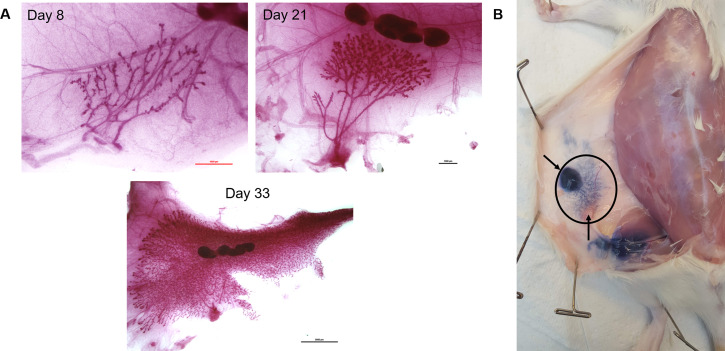
Figure 2. Rat mammary fat pad development and location.
A. Representative carmine stained whole-mount images of the fourth mammary fat pad from female Charles Rivers Sprague-Dawley rats at postnatal days 8, 21, and 33 showing the mammary ductal tree, stroma, and lymph node. Images were taken at 4x (scale bar 1,000 µm), 2x (scale bar 1,000 µm), and 0.8x (scale bar 5,000 µm) respectively. B. Trypan blue dye was injected into the nipple of the fourth mammary fat pad following euthanasia of a 25-day old female rat to indicate the location and size of the fourth mammary fat pad (inside black oval). The top arrow points to the approximate location of the nipple where dye was injected. The bottom arrow points to an intramammary inguinal lymph node. At 25 days the ductal tree has reached the lymph node.

