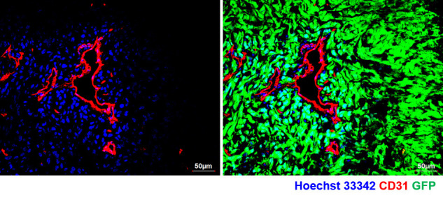
Figure 3. Confocal image of tumor section from U87MG-GFP tagged tumor xenograft retrieved post 5 min of perfusion dye (Hoechst 33342) injection and stained for BVs (CD31).

Note the spread of perfusion dye adjacent to BVs (left panel) and absent in non-perfused region of the tumor tissue. Right panel shows the presence of tumor in the whole field (GFP+ cells).
