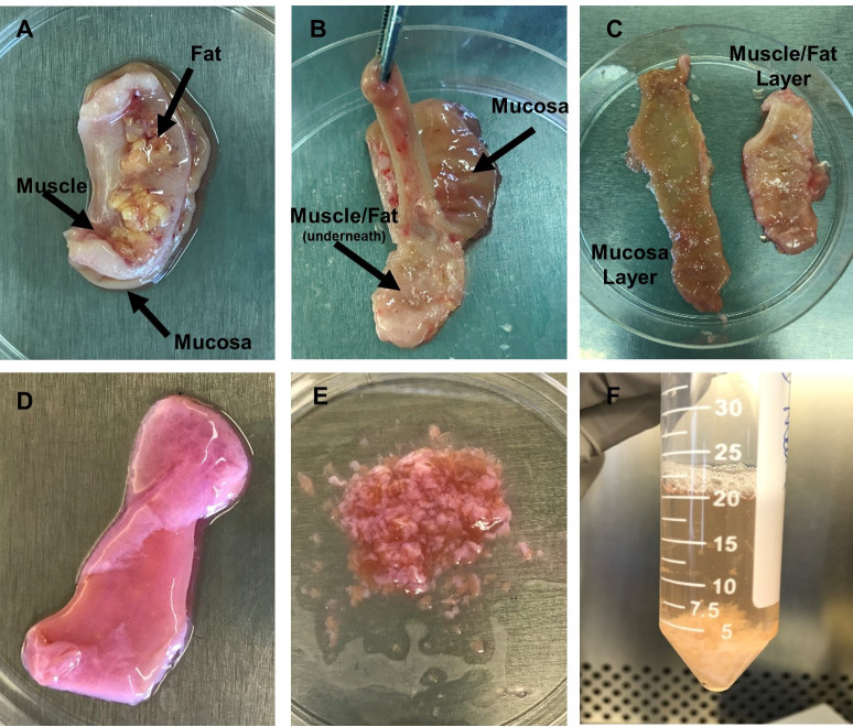
Figure 2. Images of tissue dissection and LPMC isolation.
A. Tissue is placed in a Petri dish with mucosa side down. The muscle layer and fat layer are facing upwards. B. Tissue orientation is inverted with muscle/fat layer facing downwards and mucosa layer facing upwards. Forceps are holding the mucosa layer (light brown in color) and pulling it away from the muscle/fat layer as scissors are used to separate the layers. C. Depiction of the two separated layers after dissection is complete. D. Tissue after epithelium has been removed during incubation with EDTA. Mucosa is now light pink in color. E. Minced tissue to prep for treatment with collagenase to enzymatically remove LPMCs. F. Tissue fragments and cells after incubation in collagenase. (Images by Kaylee Mickens.)

