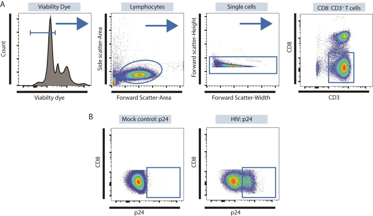
Figure 3. Identifying productively-infected LP CD4 T cells using multi-color flow cytometry.
A. Identification of CD3+ CD8- LP CD4 T cells. Viable cells are identified as Viability Dye- and then lymphocytes identified within this population based on Forward Side Scatter-Area versus Side Scatter-Area properties as shown. Doublets are excluded based on Forward Scatter-Width versus Forward-Scatter Height properties and CD3+ CD8- T cells identified within these single cells. B. Expression of p24 in CD3+ CD8- LP CD4 T cells using mock conditions as controls.

