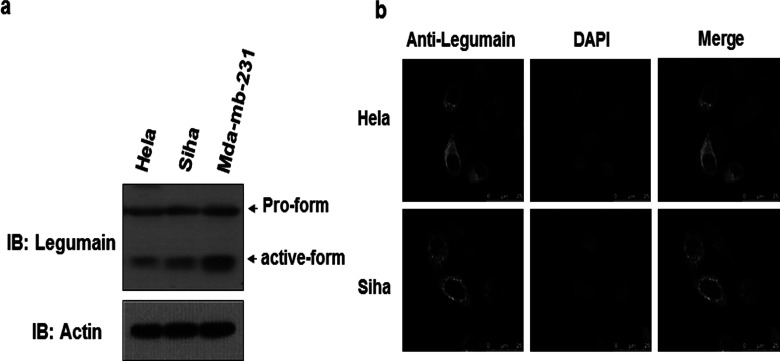
Figure 1.
Expression of legumain in cervical cancer cells. (a) Western blot of lysates of HeLa, SiHa, and Mda-mb-231 cells, probed with antibody to legumain and actin. (b) Confocal microscopy images of HeLa and SiHa cells stained with DAPI of blue fluorescence and anti-legumain of green florescence (scale bars: 25 µm).

