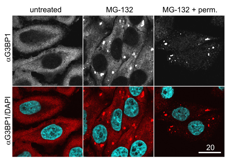
Figure 1. Immunostaining for the SG marker G3BP1 in permeabilized vs. unpermeabilized cells.
Unstressed (untreated) cells or MG-132-treated cells were fixed and stained for G3BP1 either before (MG-132) or after the cells have been permeabilized using digitonin (MG-132 + perm). Note that well-permeabilized cells (MG-132 + perm) are characterized by strong SG formation in comparison to untreated cells, but display reduced cytoplasmic staining for G3BP1 in comparison to cells that have been fixed directly after MG-132 treatment. Scale bar, 20 µm.

